


Diving in the Mediterranean, entering the water from a small cove. A few minutes later, at a few meters deep, I have an unexpected encounter: a sea urchin test. Although my dive buddy expresses annoyance by advising me to let it go, I know that this natural treasure deserves all my attention.
A gem of nature
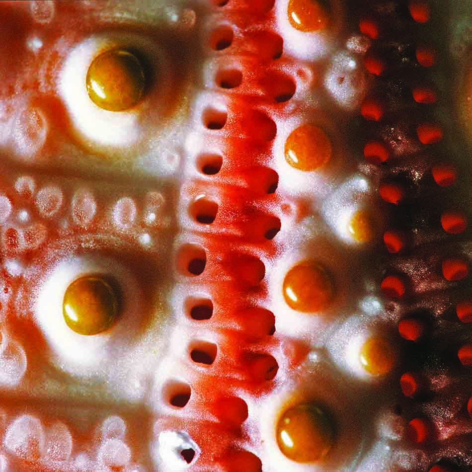
The sea urchin test that I bring home is much more than just a simple object. A true work of art from nature, it boasts a bright pink color and impressive symmetry. Its beauty lies in the details, particularly in the complex structure formed by hexagonal calcareous plates, called “assules.” These elements fascinate both architects and bioengineers, showcasing the ingenuity of nature.
Fascinating anatomy
Upon closer observation, we discover:
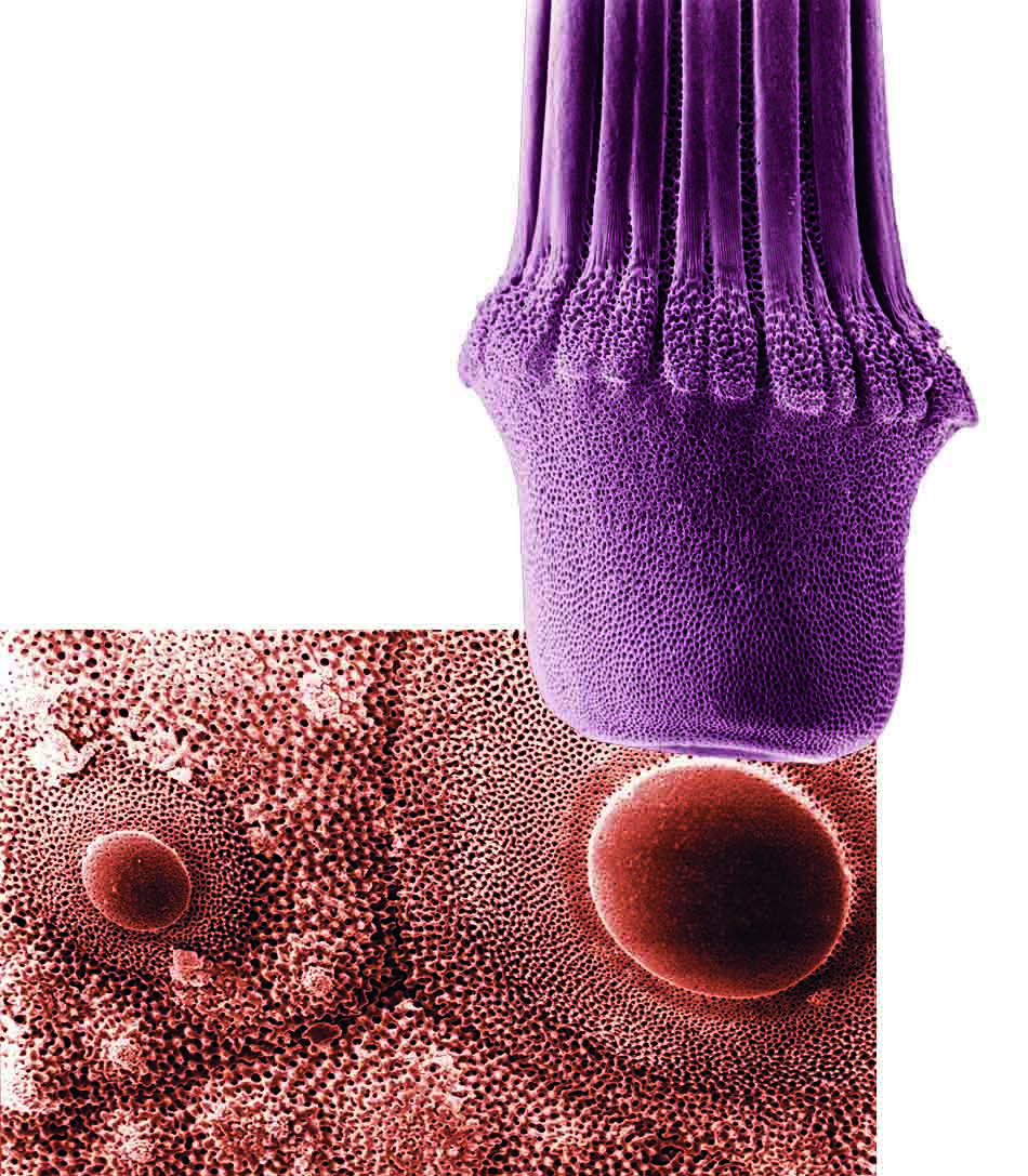
- Ambulacral zones: Five double rows pierced with small holes, through which the podia emerge. These soft and mobile tubes end with a sticky disc, allowing the sea urchin to adhere to the substrate and move. In addition to their locomotor function, these ambulacral feet play a crucial role in respiration due to their large surface area and the fineness of their epidermis.
- Hemispherical tubercles: Five broad areas are covered with small tubercles, resembling beads. Each primary tubercle is associated with a spine, or « radial, » which articulates precisely. The smaller tubercles bear finer spines, while some of them are equipped with pedicellariae. These claw-like structures help the sea urchin get rid of unwanted elements.
- Genital plates: At the top of the test, around the anal opening, are the genital plates. These allow the sea urchin to eject its eggs or sperm, depending on its sex. The madreporite, the largest of the five plates, is essential for drawing in the water necessary for the operation of its hydraulic system, which directs the podia.
A quest for knowledge
Unfortunately, my sea urchin test is incomplete, as it lacks the famous “Aristotle’s lantern,” the five-toothed mouthpart of this echinoderm. This discovery only awakens my curiosity, and I promise myself not to be distracted by the fish during my next dive. My goal will be to find a living sea urchin to explore all its secrets.
Conclusion
The sea urchin test, although often considered mundane, reveals an unexpected complexity and beauty. Every dive is an opportunity to learn and admire the richness of marine life. Next time, I will dive with a new perspective, ready to discover the hidden wonders beneath the surface.
This article, originally written by Steven Weinberg for our print edition, has been adapted for the web.
Photos: Steven Weinberg